Spectroscopic ellipsometric measurements on flakes of 2D-materials with sizes down to 1µm
Microscopic maps of thickness distribution and refractive indices from 190-1700nm
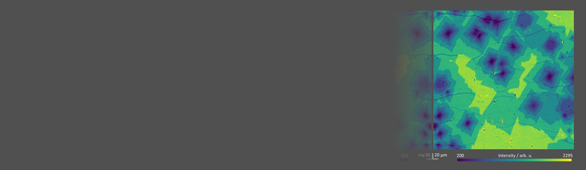
Ellipsometric contrast micrographs (ECM) enable increased contrast imaging over e.g. optical microscopy to visualize layer numbers and inhomogeneities
Non-destructive measurements on transparent substrates using knife edge illumination to prevent unwanted backside reflections
Advantages of imaging ellipsometry over other techniques
Imaging spectroscopic ellipsometry can be applied to characterize graphene and other 2D-materials. CVD grown, exfoliated and epitaxially grown flakes of 2D-materials are analyzed with the imaging ellipsometer ep4.
Imaging of monolayers on different substrates
Determination of the optical dispersion
Distinction between mono-, bi- or n-layers
Automatic flakesearch algorithm for identification of monolayers or well-defined thickness regions
Exploration of heterostructures
Quality control by detecting defects and special aspects of 2D nanoplates